GI-Miner: Advanced GI Tissue Analysis

Unlock deep insights from GI pathology

GI-Miner is a suite of tools for quantification of deep cellular and multi-cellular pathologist-friendly interpretable numerical features from gastrointestinal (GI) pathology images, supporting precise disease profiling. Leveraging state-of-the-art machine learning models, GI-Miner identifies most important cells and key histological structures, transforming complex pathology image data into rich, measurable attributes — Measurable Attributes for Pathology (MAPs) — designed for advancing biopharma research. GI-Miner is designed to support AI workflows for drug development and discovery and help improve patient stratification and enhance biomarker discovery to drive innovations in gastrointestinal disease research and practice.
Comprehensive profiling of GI tissue images
Detects key histological cells and regions, enabling quantification of pathologist-friendly histological features, providing extensive, interpretable numerical data for research.
Measurable attributes for pathology (MAPs)
Customisable feature list providing a large number of objective measurements for assessing both disease severity and response to treatment or drug candidates.
Improved tissue profiling for patient stratigication
Numerical measurements for assessing disease subtypes and risk stratification.

Precise Identification of Key Structures

Nuclei
Labelling of nuclei from key cell types: neoplastic & non-neoplastic epithelial cells, lymphocytes, neutrophils, eosinophils, plasma cells, endothelial, smooth muscle and fibroblasts.
Goblet Cells
Identification and numerical assessment of goblet cells to aid evaluation of diseases such as inflammatory bowel disease and colorectal cancer.
Tissue Type Identification
Segmentation of over 15 tissue types, including neoplastic epithelium, serrated polyps, non-neoplastic epithelium, ulceration & granulation tissue, collagen band, basophilic fringe, stroma, muscle.
Signet Ring Cells
Identification of signet ring cells, enabling assessment of signet ring cell carcinoma.
Glands and Lumen
Delineation of gland and lumen boundaries to enable morphological analyses of key histological structures.
Pigment-Laden Macrophages
Identification of pigment-laden macrophages, indicative of melanosis coli.

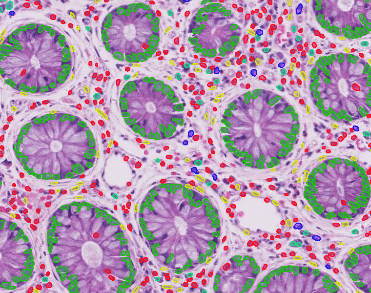
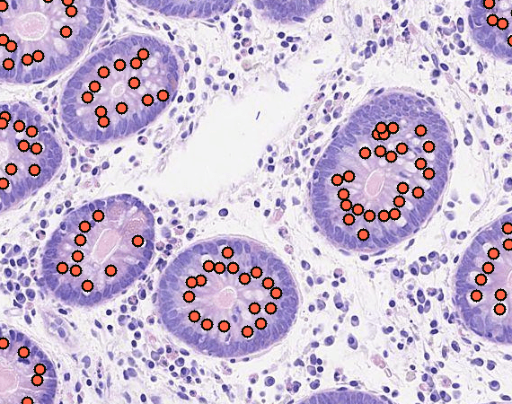
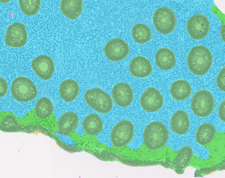
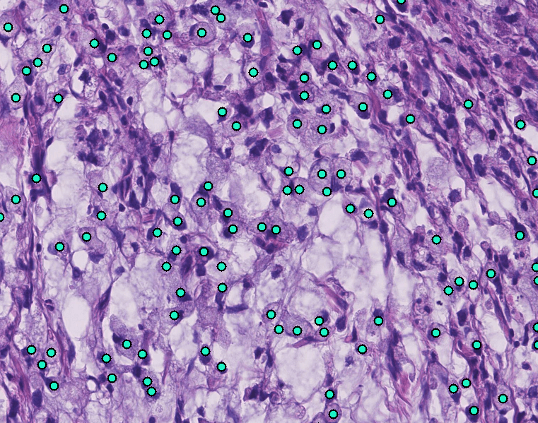
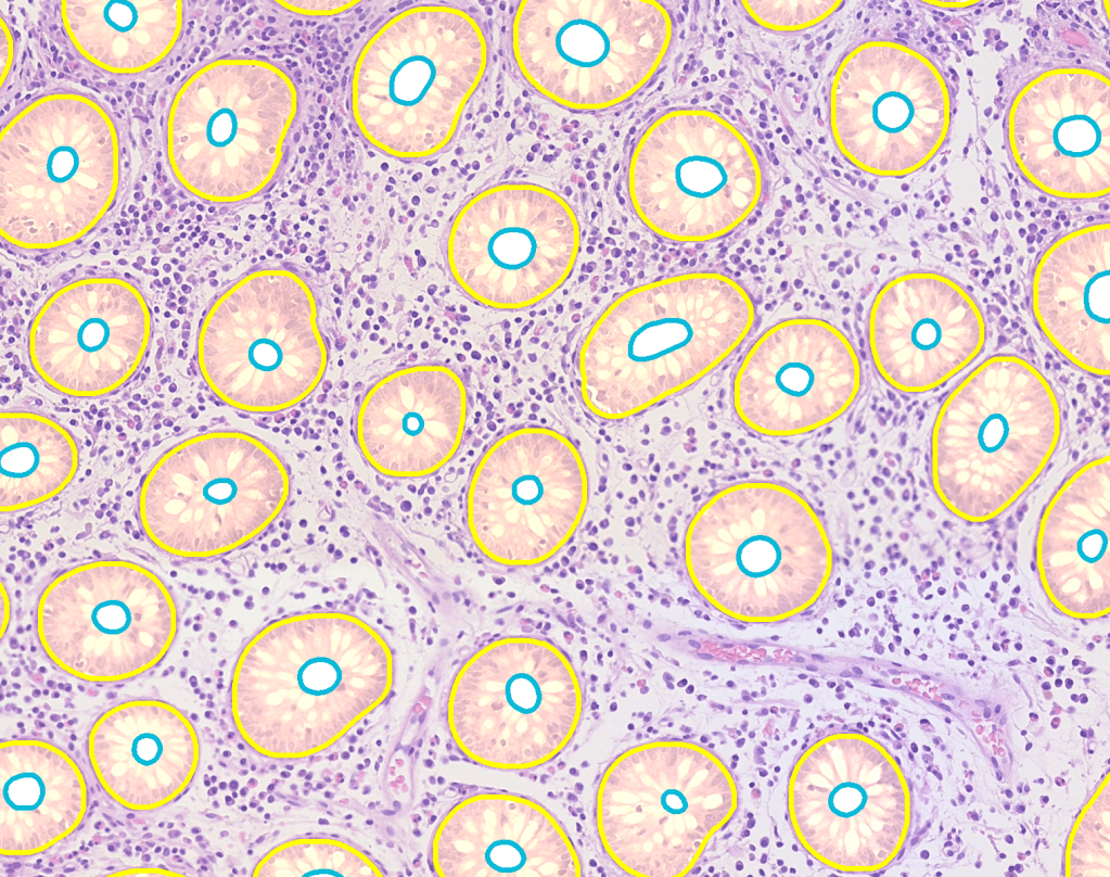
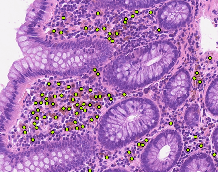
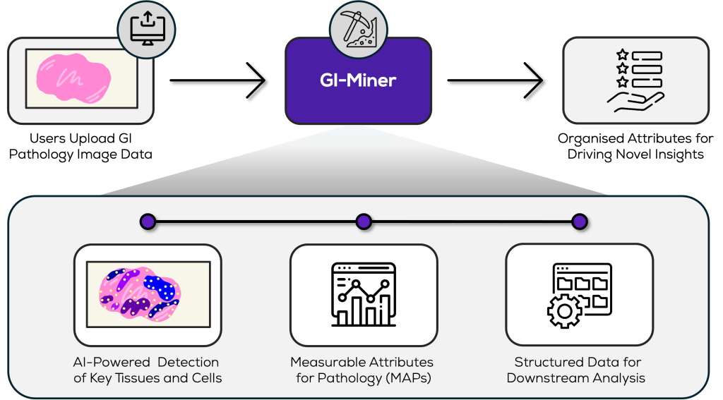
GI-Miner is a leading suite of tools for enabling the customisable measurement of a wide range of features in GI pathology images, particularly within the bowel. It supports a range of diverse applications, including the analysis of prognostic or predictive markers in colorectal cancer, assessment of inflammation severity in inflammatory bowel disease, and quantification of apoptosis in graft-versus-host disease. The suite generates a wealth of quantitative parameters from images of H&E-stained GI sections, providing valuable data for advanced research.The software suite generates structured numerical data for seamless integration into analysis pipelines, facilitating further research together with a summary report of the mined features. When paired with new technologies like spatial transcriptomics, GI-Miner enables deeper investigation into candidate cellular pathways driving the observed morphology.

Measurable Attributes for Pathology

Want to learn more about GI-Miner?